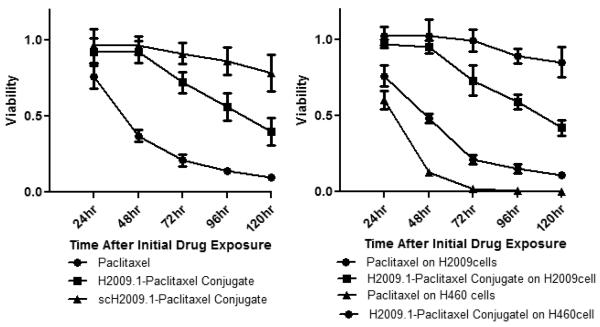
Figure 4. The H2009.1-paclitaxel conjugate displays αvβ6–selective cytotoxicity that is time dependent.
Left Panel: H2009 cells were exposed to 1 μM Taxol, H2009.1 peptide-paclitaxel (compound 3) and scH2009.1 peptide-paclitaxel for 10min, washed, and incubated further in fresh medium for different time point. The cell viability is normalized to untreated control cells. Right Panel: H2009 cells and H460 cells were exposed to 1 μM Taxol and compound 3 for 10min, washed, and incubated further in fresh medium for indicated times. The short exposure time to the conjugates is necessary to assure that the affects observed are due to the conjugate and not paclitaxel released from the peptide before internalization.

