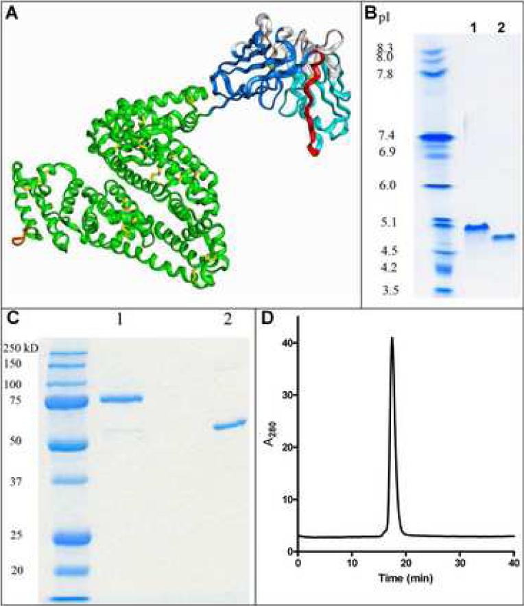
Figure 1.
Molecular model and biochemical characterization of the scFv-albumin fusion protein. A) A molecular model of the fusion protein was developed based on the crystal structures of the individual proteins. Colors are VL (light blue), VH (dark blue), CDRs (white), GS18 linker (red), HSA (green), disulfide bonds (yellow) and His6 tag (orange). The purified immunobumin (lane 1) and HSA (lane 2) were analyzed by B) isoelectric focusing gel and C) SDS-PAGE under non-reducing conditions. D) The immunobumin was also analyzed by HPLC-SEC.

