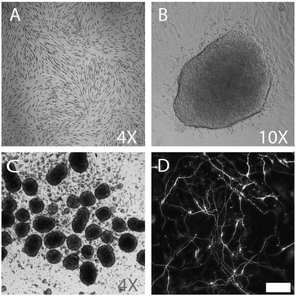
Figure 1.
IPSC induction and neural differentiation from autopsy donor-derived fibroblasts. Brightfield, phase contrast, and immunofluorescence images of (A) autopsy donor-derived dermal fibroblasts (F02AA1; Wright-Giemsa contrast stain), (B) iPSC colony arising from feeder-free conditions 21 days post transduction, (C) EBs generated from iPSC clone 2-13, and (D) neurons after 14 days of in vitro differentiation (neuron-specific beta III tubulin antibody stained). Scalebar = 50μm

