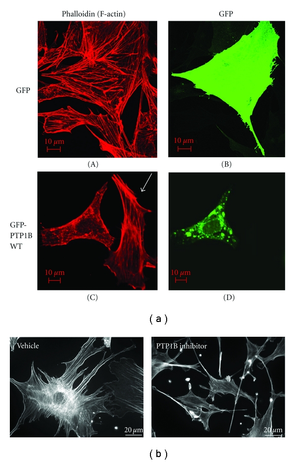
Figure 6.

Both overexpression and inhibition of PTP1B cause deranged actin cytoskeleton and cell morphology in podocytes. (a) Mouse podocytes were differentiated for 10 days and were transfected with GFP alone or GFP-PTP1B-WT. On the following day, cells were fixed, permeabilised, and stained with phalloidin to visualize F-actin. GFP transfected cells (A) or untransfected cells (C, arrow) showed well-defined stress fibers, as reported previously. Cells transfected with GFP-PTP1B-WT (C, left) showed less stress fibers and fine aggregates of F-actin. (b) Mouse podocytes were differentiated for 10 days in the presence or absence of the PTP1B inhibitor (50 μM, IC50: 4–8 μM, see Section 2). The inhibitor caused marked morphological changes with smaller cell size, collapsed appearance of cellular processes, and elongated cell shape, although the stress fibers were well preserved.
