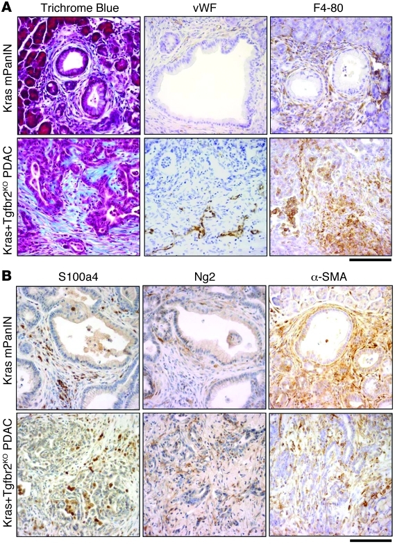
Figure 1. Stromal components in Ptf1acre/+;LSL-KrasG12D/+;Tgfbr2flox/flox PDAC and Ptf1acre/+;LSL-KrasG12D/+ mPanIN tissue.
(A) Ptf1acre/+;LSL-KrasG12D/+;Tgfbr2flox/flox PDAC stroma demonstrated prominent collagen deposition with Trichrome Blue staining and abundant macrophage and histiocyte infiltration with F4-80 immunostaining. PDAC also showed positive vascular endothelial cell staining with von Willebrand factor immunostaining, specifically in the invasion front area. Ptf1acre/+;LSL-KrasG12D/+ mPanIN stroma also showed stromal expansion to a lesser extent. (B) Fibroblast antigens (S100a4, Ng2, α-SMA) were abundantly observed in the stroma of both mPanIN and PDAC. Scale bars: 125 μm.

