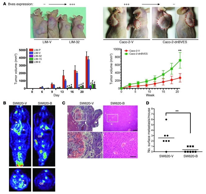
Figure 8. BVES modifies tumor growth and metastasis in athymic mice.
(A) 2.5 × 10⁶ LIM2405-P (n = 3), LIM2405-V (n = 6), Caco-2-V, Caco-2-dnBVES, or LIM2405-BVES–expressing cell lines LIM-9 (n = 6), LIM-32 (n = 3), LIM-15 (n = 3) were implanted in the dorsal flank of 8-week-old athymic nude mice. Quantification of growth rates achieved by measured tumor dimensions at the indicated intervals is presented as tumor volume (volume = [width2 × length] / 2) (average volume ± SEM. *P < 0.05, **P < 0.01, #P < 0.001, 2-way ANOVA . Splenic metastasis assay. SW620-V– or SW620-B–transfected (pooled) cells were injected into the splenic capsule (B) PET imaging (GB = Gallbladder, arrows indicate metastatic foci). (C) Histologic representative livers from indicated cell lines. (D) Metastasis quantification. **P < 0.01, Student’s t test. Small scale bar, 20 mM; large scale bar, 10 mM.

