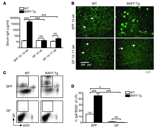
Figure 5. Commensal bacteria are necessary for hyper-IgA syndrome in BAFF-Tg mice.
(A) Serum IgA measured from SPF and GF C57BL/6 (WT) and BAFF-2 Tg mice measured by ELISA. At least 4 mice were analyzed per group (***P < 0.001). (B) Immunofluorescence staining of frozen sections of kidneys from SPF and GF C57BL/6 and BAFF-2 Tg mice stained for IgA (green). Images are representative of analysis of at least 8 mice per group (original magnification, ×100). Arrows identify glomeruli that do not exhibit IgA deposition. (C) Representative FACS profiles of IgA+ PCs in the gut LP of SPF versus GF C57BL/6 and BAFF-2 Tg mice. (D) Quantification of IgA+ PCs from C. Analysis was performed on 12-week-old BAFF-2 Tg mice versus C57BL/6 mice. Note that BAFF levels in GF BAFF-2 Tg mice were approximately equal to those observed for SPF BAFF-Tg mice (1,733.2 ± 660.8 pg/ml; n =12). LPL, LP lymphocyte. Bars in A and D represent the mean ± SEM. *P < 0.05, ***P < 0.001.

