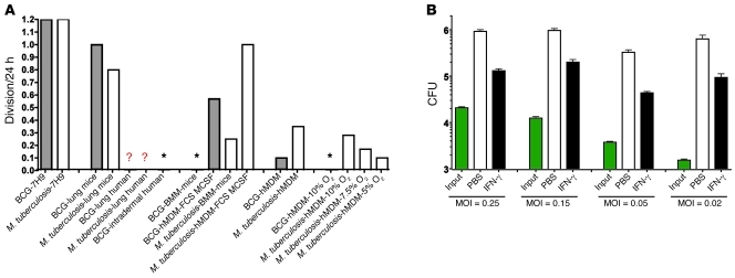
Figure 8. Approximate net number of mycobacterial divisions in mice, in humans, and in their macrophages.
(A) Approximate net mycobacterial cell divisions per 24 hours for BCG (gray bars) or M. tuberculosis (white bars) in diverse settings. hMDM, human MDMs prepared under the most effective conditions illustrated in A; hMDM-MCSF-FBS, conventionally cultured human hMDM, hMDMs after about 1 week in 10% FCS with M-CSF in 20% O2; lung mice, logarithmic growth phase in the lungs of immunocompetent mice over the first 3 weeks following low-dose M. tuberculosis aerosol infection. Lung human, lungs in humans. Lung mice, lungs in mice. Asterisks, no net increase; mycobacteria are killed. Question mark indicates that no data are available, but it is expected that after a brief period of replication, there is no further net increase in more than 90% of immunocompetent people for M. tuberculosis and 100% for BCG. Figures for mycobacteria in host cells in vitro are based on periods of 3 weeks for BCG in hMDMs, 2 weeks for M. tuberculosis in hMDMs, and 1 week for BCG and M. tuberculosis in moBMMs. (B) Effect of MOI on the number of net divisions of M. tuberculosis in MDMs. MDMs were differentiated without exogenous cytokines in 10% O2 for 14 days, stimulated with IFN-γ (3 ng/ml) or not on day 14, infected with M. tuberculosis at the indicated MOI on day 16, and lysed 2 weeks later for determination of CFU (log10 scale). Results are representative of those with MDMs from 2 donors tested in 4 independent experiments.

