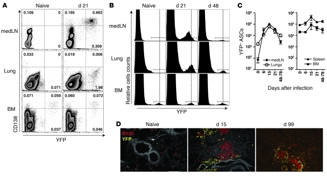
Figure 1. Kinetics of ASCs during influenza virus infection.
(A) Naive and influenza virus PR8–infected BLIMP-1–YFP mice (day 21 after infection) were analyzed for YFP+CD138+ ASCs in lung-draining medLN, lungs, and BM. Cells expressing CD4, CD8, and CD11b were excluded. Dot plots with percentages shown are representative of n = 3–5 mice from at least 2 independent experiments. (B) Level of YFP expression in ASCs from medLN, lungs and BM isolated from naive and influenza-infected BLIMP-1–YFP mice. Histograms are based on CD4–CD8–CD11b– cells and representative of n = 5–7 mice. (C) Kinetics of YFP+ ASCs during influenza virus infection. Data with mean ± SEM are representative of n = 3–5 mice per time point. (D) Immunofluorescence of lungs from naive and infected BLIMP-1–YFP mice stained for B220 (red). Original magnification, ×20, Scale bar: 100 μm.

