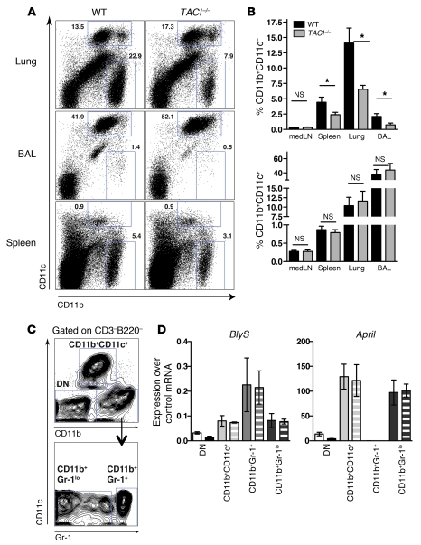
Figure 7. Reduction of BLyS- and APRIL-expressing CD11b+CD11c– cells in lungs of TACI–/– mice after influenza virus infection.
(A) Flow cytometry plots with percentages of CD11b+CD11c+ and CD11b+CD11c– cells in lungs, BAL, and spleen isolated from WT or TACI–/– mice at day 34 p.i. (B) Frequency of CD11b+CD11c– and CD11b+CD11c+ cells in various organs of WT and TACI–/– mice at day 34 p.i. Data with mean ± SEM combine the results of 3 independent experiments (n = 8 mice/group). (C) Identification of cell subsets in lungs of mice based on expression of CD11b, CD11c, and Gr-1 used for purification by FACS. DN, double negative (CD11b–CD11c–). (D) Real-time PCR analysis of BLyS and April mRNA expression in cell subsets sorted from lungs as shown in C of naive mice (solid bars) and mice 3–4 weeks p.i. Data with mean ± SEM are from 2 independent experiments. *P < 0.05.

