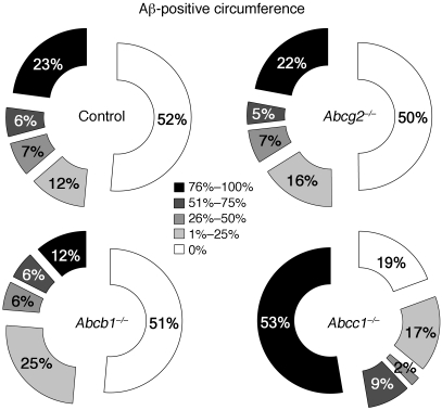
Figure 5. Aβ deposition is enhanced in the walls of brain microvessels in APPdt×Abcc1–/– mice.
The graphs present the proportion of meningeal vessel walls that were affected by different degrees of Aβ deposition (Aβ-positive circumference). Assessment of APPdt mice deficient in ABCG2 or ABCB1, respectively, revealed no differences in CAA relative to APPdt controls. In contrast, 53% of blood vessels were severely affected by CAA (>75% of the circumference is decorated with Aβ) in APPdt×Abcc1–/– mice versus 23% in APPdt controls. n = 3.

