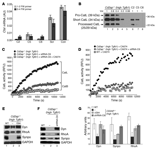
Figure 3. Cytosolic CatL activity regulates actin cytoskeleton in Cd2ap–/– cells.
(A) Ctsl mRNA levels, determined by RT-PCR, in high-Tgfb1 Cd2ap–/– podocytes infected with different shRNA constructs (C2, C5, C6) downregulating endogenous CatL. Con, high-Tgfb1 Cd2ap–/– podocytes not infected with lentiviruses; Scr, cells infected with lentiviruses expressing a scrambled oligo. (B) CatL levels in high-Tgfb1 Cd2ap–/– podocytes infected with lentiviruses expressing different shRNA constructs to downregulate CatL at relative volumes as indicated. (C) Time course of CatL activity in high-Tgfb1 Cd2ap–/– podocytes and in high-Tgfb1 Cd2ap–/– podocytes infected with lentiviruses to downregulate CatL in the absence and presence of CA074 at neutral pH. (D) Time course of CatL activity in the presence of CA074 in high-Tgfb1 Cd2ap–/– podocytes, high-Tgfb1 Cd2ap–/– podocytes infected with lentiviruses to downregulate CatL, and WT podocytes at neutral pH. (E and F) Protein levels in WT cells, high-Tgfb1 Cd2ap–/– podocytes, and high-Tgfb1 Cd2ap–/– podocytes treated with E64 or infected with lentiviruses to downregulate CatL (shRNA-C6). Dyn, dynamin; Synpo, synaptopodin. GAPDH was used as a loading control. (G) Quantitation of protein levels from Western blots in E and F.

