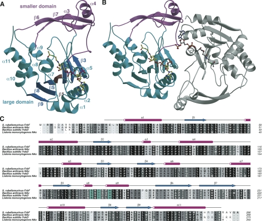
FIGURE 3.
Overall structure and multiple sequence alignment. A, ribbon diagram showing the overall structure of the FrbF monomer in complex with acetyl-CoA (in yellow ball-and-stick). The acetyl-CoA-binding large domain is shown in cyan, and the smaller domain is shown in pink. B, ribbon diagram showing the structure of the FrbF dimer with the second molecule colored in gray. C, structure-based sequence alignment of FrbF along with the structural homologs B. anthracis N-acetyltransferase (NAc), B. subtilis YokD, and the aminoglycoside N-acetyltransferase from L. monocyotogenes. Sequence conservation is shaded such that the residues that show the highest conservation are darker. Residues colored in cyan are active site general acid/base catalysts.

