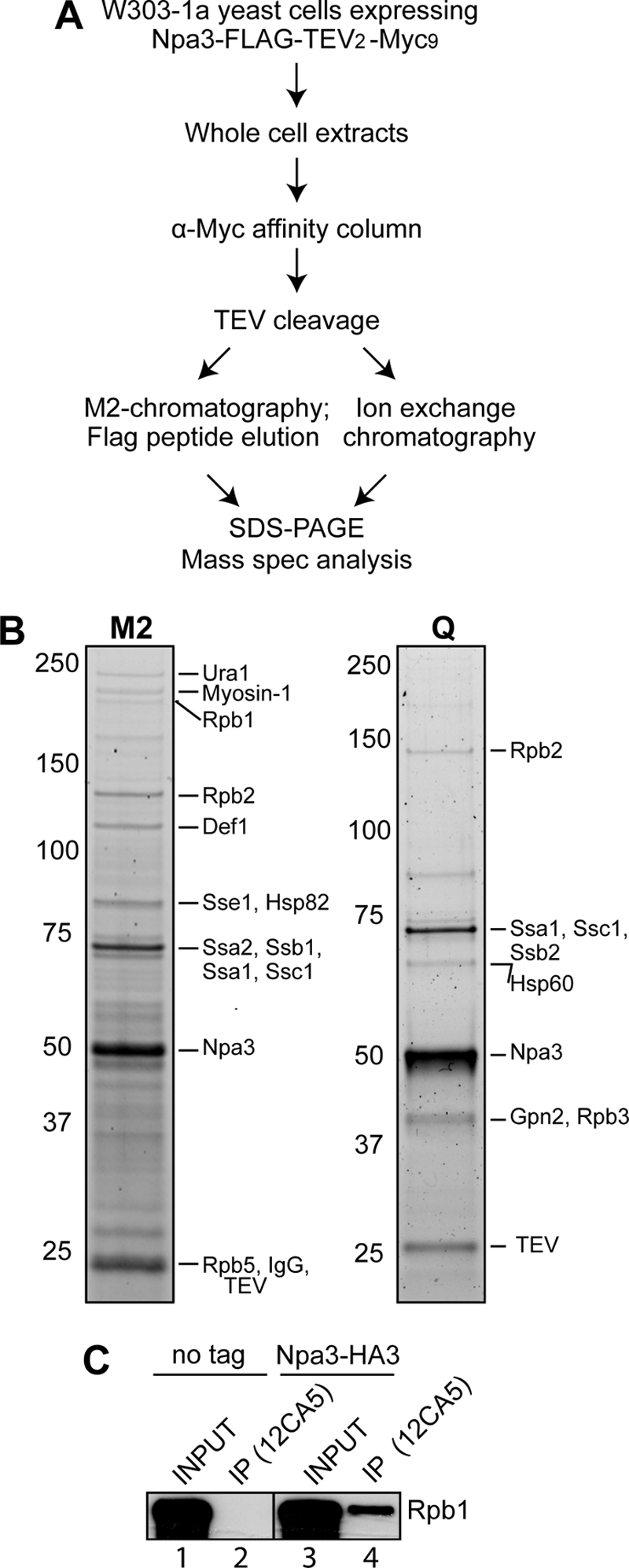
FIGURE 1.

RNAPII co-purifies with Npa3. A, outline of the Npa3 purification procedure. Mass spec analysis, mass spectrometric analysis. B, eluates from M2-agarose (M2) and a HiTrapQ FF column (Q) (peak fraction) were separated by 4–12% SDS-PAGE and stained with SYPRO Ruby. Positions of proteins identified by mass spectrometric analysis are indicated on the right, and marker protein migration is indicated on the left. C, immunoprecipitation (IP) from whole cell extracts from a strain expressing endogenous Npa3 with a C-terminal 3×HA tag (Npa3-HA3) or an untagged control strain (no tag), with anti-HA (12CA5) antibody. Rpb1 was detected by Western blotting with a mixture of 4H8 and 8WG16 antibodies.
