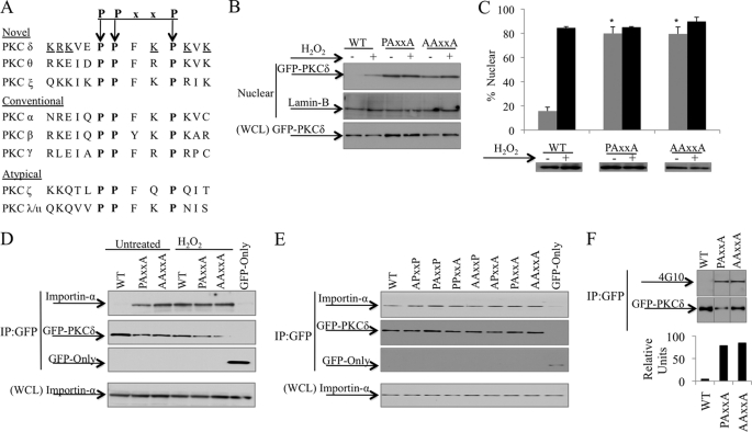
FIGURE 4.
The PPxxP motif facilitates retention of PKCδ in the cytoplasm. A, a conserved PPxxP motif (boldface) overlaps the bipartite NLS (underlined) of PKCδ. B, 293T cells were transfected with pGFP-WT-PKCδ (WT), pGFP-PAxxA-PKCδ (PAxxA), or pGFP-AAxxA-PKCδ (AAxxA) and were left untreated or treated with 5 mm H2O2 for 30 min. Nuclear fractions and WCL were separated by SDS-PAGE, and Western blots were probed with an anti-GFP antibody. Lamin-B was used as a loading control for nuclear fractions. C, ParC5 cells were transfected with pGFP-WT-PKCδ (WT), pGFP-PAxxA-PKCδ (PAxxA), or pGFP-AAxxA-PKCδ (AAxxA) and either left untreated (gray bars) or treated with 5 mm H2O2 (black bars) for 1 h. Nuclear localization of GFP-tagged proteins was analyzed by fluorescence microscopy. The Western blot shows protein expression of the different constructs. Asterisks indicate a statistically significant difference from untreated WT (Student's t test; p < 0.002). D, 293T cells were transfected with pGFP, pGFP-WT-PKCδ (WT), pGFP-PAxxA-PKCδ (PAxxA), or pGFP-AAxxA-PKCδ (AAxxA) and were either left untreated or treated with 5 mm H2O2 for 30 min. E, 293T cells were transfected with the indicated pGFP2-PPxxP constructs or pGFP. D and E, upper panels, the GFP-tagged proteins were immunoprecipitated from WCL using an anti-GFP antibody and assayed by Western blot for endogenous importin-α interaction and the amount of GFP-tagged proteins immunoprecipitated (IP). Lower panels, WCL was assayed by Western blot to show the amount of importin-α present in the lysate. F, 293T cells were transfected with pGFP-WT-PKCδ (WT), pGFP-PAxxA-PKCδ (PAxxA), or pGFP-AAxxA-PKCδ (AAxxA). The GFP-tagged proteins were immunoprecipitated from WCL using an anti-GFP antibody and assayed by Western blot for total PKCδ tyrosine phosphorylation (4G10) and the amount of GFP-tagged immunoprecipitated. The graph represents densitometric analysis of the above Western blots quantifying the amount of PKCδ total tyrosine phosphorylation relative to protein levels. For all panels, each experiment was repeated three or more times; a representative experiment is shown.

