FIGURE 1.
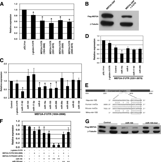
The 3′-UTR of the Mef2A gene mediates its repression. A, luciferase reporters containing the 3′-UTRs of the mouse Mef2A gene were transfected into the 293T cells, and luciferase activity was measured 48 h later. Results were presented as relative luciferase activity in which the control was assigned a value of 1. The pGL-3-luciferase and the β-globin-3′-UTR-luciferase reporters were used to serve as controls. Data represent the mean + S.D. from at least three independent experiments in triplicate. *, p < 0.05. B, equal amount of FLAG-tagged MEF2A expression plasmids, which contains or does not contain its 3′-UTR were transfected into 293T cells. Protein expression level was measured by Western blot. β-Tubulin was used as a loading control. C, a luciferase reporter directed by the full-length MEF2A-3′-UTR(1834–2898) was co-transfected with indicated miRNA expression plasmids into the 293T cells, and luciferase activity was measured 48 h later. Results were presented as relative luciferase activity in which the control was assigned a value of 1. The pGL-3-luciferase (control) and the β-globin-3′-UTR-luciferase reporters were used to serve as controls. Data represent the mean + S.D. from at least three independent experiments in duplicate. *, p < 0.05. D, a luciferase reporter directed by a short MEF2A-3′-UTR(2251–2679) was co-transfected with indicated miRNA expression plasmids into the 293T cells, and luciferase activity was measured 48 h later. Results were presented as relative luciferase activity in which the control was assigned a value of 1. The pGL-3-luciferase and the β-globin-3′-UTR-luciferase reporters were used to serve as controls. Data represent the mean + S.D. from at least three independent experiments in duplicate. *, p < 0.05. E, schematic diagram of the mouse Mef2A 3′-UTR and the sequence alignment with miR-155. F, luciferase reporters containing the 3′-UTRs of the mouse Mef2A gene (full-length nt 1834–2898 or the short form nt 2251–2679) were co-transfected with increasing amount of miR-155 or a mutant miR-155 (miR-155-mut) into the 293T cells, and luciferase activity was measured 48 h later. Results were presented as relative luciferase activity in which the control was assigned a value of 1. The β-globin-3′-UTR-luciferase reporter was used to serve as controls. Data represent the mean + S.D. from at least three independent experiments in triplicate. *, p < 0.05. G, FLAG-tagged MEF2A expression plasmids, which contain its 3′-UTR, were co-transfected with miR-155 or a mutant miR-155 into 293T cells. Protein expression level was measured by Western blot using an anti-FLAG antibody. β-Tubulin was used as a loading control.

