FIGURE 4.
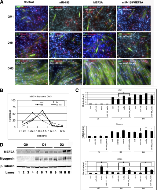
miR-155 inhibits myoblast differentiation which is suppresses by MEF2A. A, immunohistology of C2C12 cells treated with control, miR-155, Ad-MEF2A, or both miR-155 and Ad-MEF2A. Cells were induced to differentiate at indicated dates and were stained with antibodies that recognize striate muscle MHC (green) and myogenin (red). DAPI stains nuclei. GM1, growth medium day 1; DM1, differentiation medium day 1; DM3, differentiation medium day 3. B, quantitative analyses of cell size of MHC positive myoblast and myotubes in C2C12 cells treated with control, miR-155, MEF2A, or both miR-155 and MEF2A. The distribution of myoblast and myotube size was presented as percentage. C, quantitative real-time PCR analyses of gene expression in C2C12 cells treated with control, miR-155, MEF2A, or both miR-155 and MEF2A. Result was presented as relative expression level in which the control was assigned a value of 1. Data represent the mean + S.D. from at least three independent experiments. *, p < 0.05. G0, growth medium day 0; D1, differentiation medium day 1; D3, differentiation medium day 3. D, Western blot analyses of protein expression levels in C2C12 cells treated with control (contl), miR-155, MEF2A, or both miR-155 and MEF2A. D2, differentiation medium day 2.

