FIGURE 1.
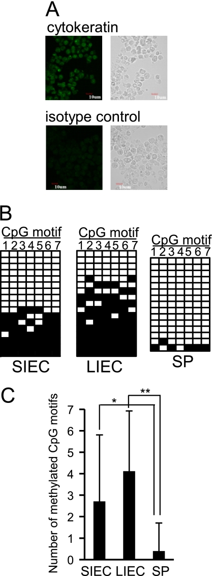
The 5′ region of the TLR4 gene is highly methylated in IECs in vivo. A, expression of cytokeratin, a marker of epithelial cells, in SIECs and LIECs prepared from mice was confirmed by immunostaining. Cells were stained with FITC-conjugated anti-pan cytokeratin antibody (upper panels) or FITC-conjugated isotype control antibody (lower panels). Left panels show fluorescent images under confocal laser scanning microscopy, and right panels show transmission images under phase-contrast microscopy. A representative staining pattern of SIECs is shown. Similar staining patterns were obtained for LIECs. B, methylation frequencies of CpG motifs existing in the 5′ region of the TLR4 gene were compared among mouse SIECs, LIECs, and splenic cells (SP). DNA methylation of 7 CpG motifs in the 5′ region (nucleotides −102/+202) was analyzed by the bisulfite conversion reaction. The modified DNA from 6 mice was amplified by PCR, cloned, and sequenced for 15–17 independent clones. A methylation pattern of 7 CpG motifs (1–7) for each clone is shown in the order of methylation frequency. Filled squares indicate methylated CpG motifs, whereas open squares indicate unmethylated CpG motifs. C, mean numbers of methylated CpG motifs ± S.D. are represented in the graph. * and **, significantly different (*, p < 0.05; **, p < 0.0001).

