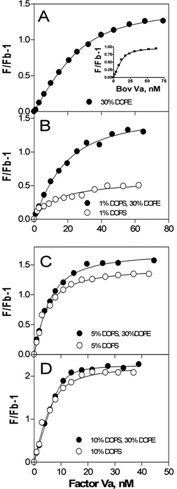
FIGURE 3.

Binding of factor Va to phospholipid vesicles. Samples contained 150 mm NaCl, 20 mm Tris, pH 7.4, and 0.6 μm labeled DOPC:DOPS:DOPE:dansyl-PE vesicles of varying compositions: 70:0:27.5:2.5 (A), 69:1:27.5:2.5 and 96.5:1:0:2.5 (B), 65:5:27.5:2.5 and 92.5:5:0:2.5 (C), and 60:10:27.5:2.5 and 87.5:10:0:2.5 (D). Fluorescence measurements (F) were made as described under “Experimental Procedures” after incremental addition of bovine factor Va at 25 °C. The lines represent the best fit of data in one representative experiment. The inset in A shows the binding of bovine Va to egg PE:egg PC (20:80) membranes.
