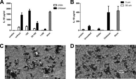
FIGURE 3.
The effect of particle size on inflammasome activation. A, chitosan and chitin preparations prepared as in Fig. 1 were sonicated and then size-fractionated through 100-μm and 20-μm filters. BMMΦ (1 × 10⁵/well) were primed with LPS and then stimulated with chitosan or chitin particles (1 mg/ml) that were left unfractionated (unfract) or size-fractionated as indicated. IL-1β was analyzed by ELISA. Data are means ± S.E. of three independent experiments, each performed in triplicate. p < 0.001 comparing unfractionated chitosan to 20–100 chitosan and >100 chitosan fractions, and between the <20 chitosan fraction and the 20–100 and >100 chitosan fractions, analyzed by two-way ANOVA. B, LPS-primed BMMΦ (1 × 10⁵/well) were left unstimulated (Unstim) or incubated for 6 h with the indicated size and type of beads (1 mg/ml). Alum (1 mg/ml) served as a positive control. Supernatants were analyzed for IL-1β by ELISA. Data are means ± S.E. of three independent experiments, each performed in triplicate. p < 0.01 comparing 3-μm chitosan beads and 50-μm chitosan beads by two-way ANOVA. Shown are representative photomicrographs of BMMΦ following 30-min incubation with 3-μm chitin-coated (C) and chitosan-coated (D) beads demonstrating robust phagocytosis of both types of glycan-coated beads.

