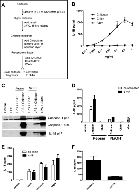
FIGURE 4.
Effect of pepsin digestion of chitosan on inflammasome activation. Following the procedure outlined in A, chitosan was digested with pepsin and then half was converted to chitin. B, dose curve of the pepsin-treated chitin and chitosan-stimulating BMMΦ (1 × 10⁵/well) after they were primed for 3 h with 100 ng/ml LPS. Data are means ± S.E. of four independent experiments, each performed in triplicate. p < 0.01 comparing chitin and chitosan at any concentration ≥ 0.1 mg/ml as analyzed by unpaired t test. C, BMMΦ (1.5 × 10⁶/well) were primed for 3 h with 100 ng/ml LPS and then stimulated with alum (0.1 mg/ml) or chitin and chitosan derived from the procedure outlined in Fig. 4A (pepsin) or the procedure outlined in Fig. 1A (NaOH). Supernatants were then collected and analyzed for caspase-1 and IL-1B by immunoblot. Caspase-1 p20 and IL-1B p17 represent the mature forms and indicate an active inflammasome, whereas caspase-1 p45 is an inactive proform of caspase-1. D, BMMΦ (1 × 10⁵/well) were primed as in B and then stimulated with alum or chitin and chitosan derived from the procedure outlined in Fig. 4A (pepsin) or the procedure outlined in Fig. 1A (NaOH). The chitin and chitosan preparations were left unsonicated (no sonication) or sonicated for 5 min (5 min). All stimuli were added at a concentration of 0.1 mg/ml. Supernatants were analyzed by ELISA for IL-1β. Data are means ± S.E. of three independent experiments, each performed in triplicate. p < 0.001 comparing no sonication and 5-min sonication of pepsin chitosan by two-way ANOVA. E, BMMΦ (1 × 10⁵/well) were primed as in B. Two hours later, wells either received 0.1 mg/ml chitin or were left without chitin treatment (no chitin). One hour later, cells were left unstimulated (unstim) or stimulated for 6 h with alum (0.1 mg/ml), or chitosan (0.1 mg/ml), or 1 h with nigericin (2.5 μm). Supernatants were analyzed by ELISA for IL-1β. Data are mean ± S.E. of two independent experiments, each performed in triplicate. F, BMMΦ (1 × 10⁵/well) were primed as in B. Insoluble suspended chitosan and chitosan that had been solubilized in acetic acid were diluted in media and added to cells. Supernatants were analyzed by ELISA for IL-1β. Data are means ± S.E. of two independent experiments, each performed in triplicate. p < 0.01 comparing insoluble with soluble chitosan by two-tailed unpaired t test.

