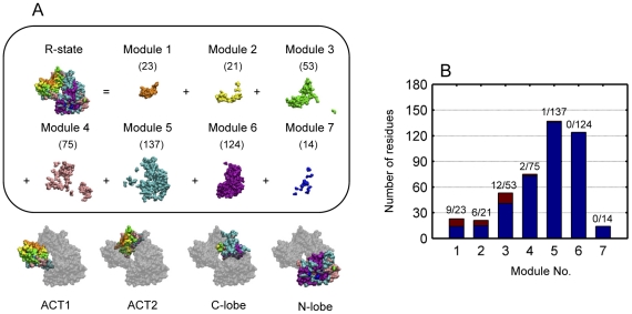
Figure 5. Protein dynamical modules of E. coli aspartokinase III and distribution of mutation sites.
(A) Protein dynamical modules are obtained at a time interval of 0.1 ps. Modules are differently colored and numbers of residues within each dynamical module are given in brackets. (B) Numbers of residues within each protein dynamical module (colored in blue and brown, denominator) and distribution of mutation sites predicted by integrating molecular dynamics and co-evolutionary analysis [23] among each module (colored in brown, numerator).

