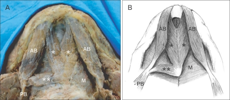
Fig. 1.
Photograph (A) and schematic drawing (B) showing the accessory bellies of the digastric muscle. Two accessory bellies (*) merged and some parts are attached to the mylohyoid raphe. Right inferior portion continued as 3rd accessory belly (**) and inserted right intermediate tendon. AB, anterior belly; M, mylohyoid muscle; PB, posterior belly.

