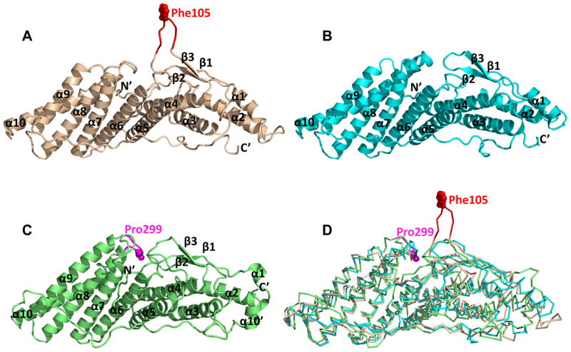
Figure 2. Structural superposition of the three Bro1 domains.
The Bro1 domains of Alix, HD-PTP and Brox are shown as wheat, cyan and green ribbons, respectively in (A–C), and superimposed as Cα traces in (D). Residue Phe105 of Alix is shown as red spheres in (A) and (D), and reidue Pro299 of Brox is shown as magenta spheres in (C) and (D). The secondary structures as well as the N- and C-termini of the three Bro1 domains are marked in (A–C). See also Figure S1.

