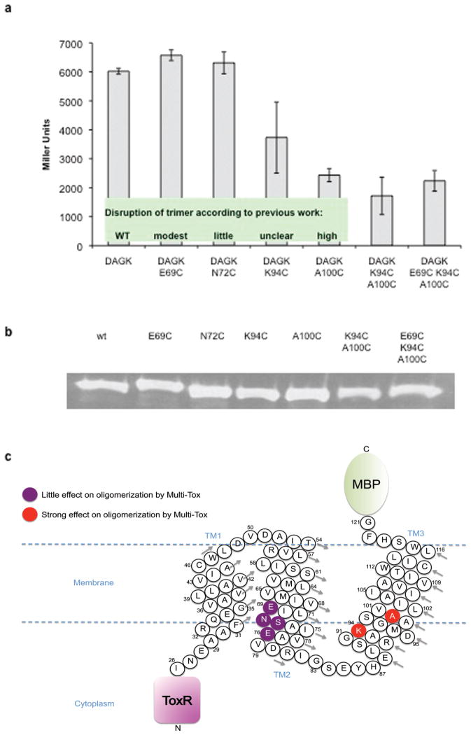
FIGURE 2.
(a) Analysis of key residues in the DAGK TMD by Multi-Tox. Error bars represent the standard deviation of three replicates, each measured in quadruplicate. Inset: classification of DAGK mutants for disruption of trimerization as previously reported [15]; (b) Western blots of protein expression for DAGK TMD; (c) Cartoon representation of the MBP-DAGK-ToxR Multi-Tox chimeric protein, summarizing the DAGK residues investigated. DAGK residues are numbered according to the wild-type unmodified DAGK protein. Arrows indicate direction of numbering for each helix turn.

