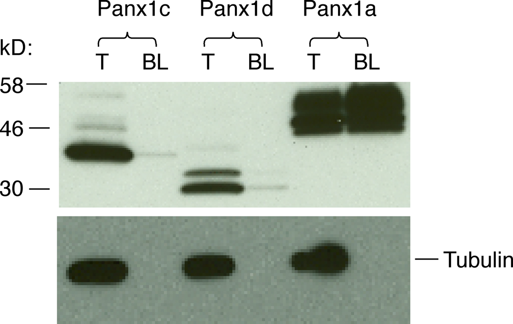
Fig. 6.
The biotin labeling analysis of the cell surface localization of Panx1 isoforms. HEK293 cells were transfected with N-terminal FLAG-tagged Panx1 constructs and 48 h later labeled with the membrane impermeable Sulfo-NHS-SS-Biotin. Total cell lysates (T) and biotin-labeled plasma membrane protein fractions (BL) were probed with anti-FLAG HRP-labeled antibody (top panel) and the same membranes were reprobed with anti-alpha tubulin antibody (controls, bottom panel). Note that only Panx1a was successfully biotinylated.

