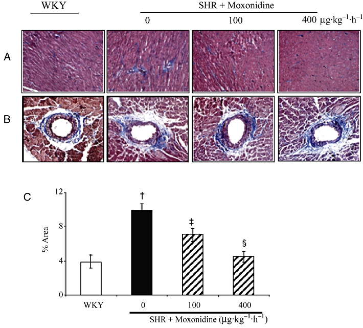
Figure 1.

Representative photomicrographs of Masson's Trichrome stained heart sections of spontaneously hypertensive rats (SHR) and Wistar-Kyoto (WKY) rats with and without moxonidine treatment. (A) Interstitial, (B) perivascular collagen deposition (in blue) and (C) percentage of interstitial and perivascular collagen deposition in total area. n = 4–6 rats per group; †P < 0.01 versus WKY; ‡P < 0.05 versus vehicle; §P < 0.01 versus vehicle. Magnification 200×.
