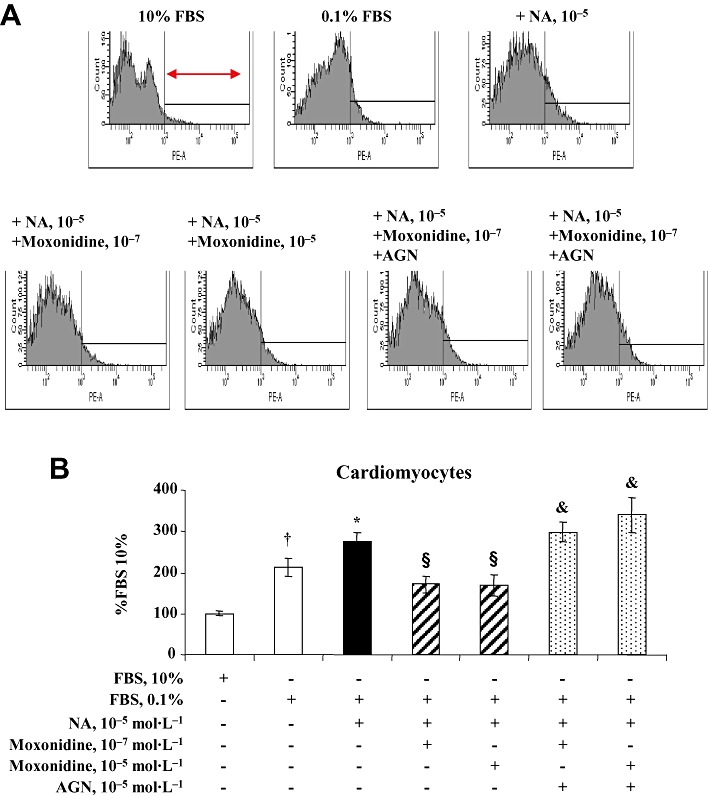
Figure 5.

(A) Representative flow cytometry and propidium iodide staining depicting total mortality of neonatal rat cardiomyocytes in culture. Cells on the right (arrow) represent the percentage of cell death out of total number of cells measured. (B) Bargraph represents cardiomyocyte mortality after 48 h incubation in Dulbecco's modified Eagle's medium (DMEM) containing 10% fetal bovine serum (FBS), and in conditions of starvation (DMEM + 0.1% FBS) alone or in addition to noradrenaline (NA) without and with co-incubation with moxonidine at 10−7 and 10−5 mol·L−1, without and with and I1-receptor antagonist, AGN 192403 (AGN) at 10−5 mol·L−1. Data presented as per cent FBS 10%. n = 8–12 wells per treatment, from five independent cultures. †P < 0.01 versus 10% FBS; *P < 0.05 versus 0.1% FBS; §P < 0.01 versus NA; &P < 0.05 versus corresponding NA + moxonidine.
