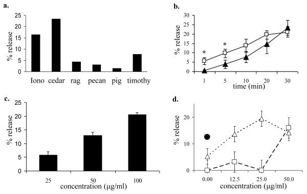
Figure 1.
Mast cell serotonin release induced by pollen extract.
a. 3H-serotonin release induced by pollens extracted in NH4HCO3 buffer. Total protein content = 100 μg/ml. Iono = ionomycin (1 μM), cedar = mountain cedar, rag = ragweed, pig = pigweed, and timothy = timothy grass.
b. 3H-serotonin release was determined in RBL-2H3 cells at 1, 5, 10, 20, and 30 minutes after stimulation with cedar pollen extract (open boxes; □) or 1 μM ionomycin (filled triangles; ▲). Data represent the means of 3 independent experiments, * indicates significant differences between ionomycin and cedar extract ( p< .05).
c. 3H-serotonin release was determined in RBL-2H3 cells after incubation with 25, 50, 100, or 200 μg/ml cedar pollen extract for 30 minutes. The results are expressed as mean ± SD (n = 3 separate experiments, each experiment performed in duplicate).
d. 3H-serotonin release in RBL-2H3 cells after exposure to pollen extract plus DNP-BSA 1 ng/ml (open triangles; △) or pollen extract alone (open boxes; □). DNP-BSA 50 ng/ml (filled circle; ●).

