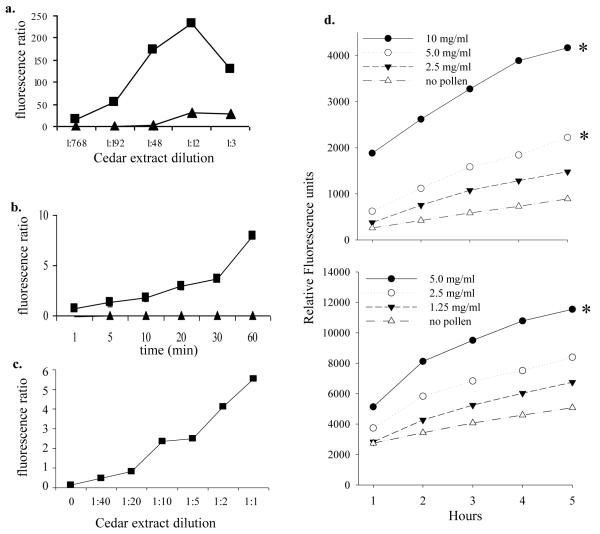
Figure 2.
ROS generation by pollen extract
a. Fluorescence of cedar pollen extracts incubated with DCFH-DA for 30 min. Cedar pollen was extracted with either NH4HCO3 (filled boxes; ■) or PBS (filled triangles; ▲).
b. DCF fluorescence of RBL-2H3 cells stimulated with 100 μg/ml pollen extracted in NH4HCO3 buffer (■) or 1 μM ionomycin (▲).
c. DCF fluorescence of RBL-2H3 cells incubated with dilutions of pollen extracts (NH4HCO3 buffer) for 30 min. The specific protein content of pollen extracts was not determined in this experiment, but in subsequent. In general, extracts contained approximately 200-300 μg/ml total protein.
d. DCF fluorescence of HMC-1 cells (top, mean of 2 experiments) or RBL-2H3 cells (bottom, single experiment) incubated with dilutions of mountain cedar pollen grains. Data are expressed as relative fluorescence units with the baseline fluorescence of each condition subtracted from subsequent measures at 1-5 hours. Pollen grains in buffer alone showed high baseline levels of autofluorescence that did not change over time (data not shown). Significant differences from the “no pollen” controls are indicated with an asterisk (*).

