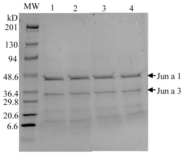
Figure 3.

Protein profile of cedar pollen extracts. Cedar pollen extracted in NH4HCO3 (lane 1); NH4Cl (lane 2); NaHCO3(lane 3); or PBS (lane 4) was analyzed by SDS-PAGE stained with Coomassi blue. The major allergens, Jun a 1 (43 kDa) and Jun a 3 (30kDa) (arrows), migrated slightly slower on the gel likely due to carbohydrate moieties.
