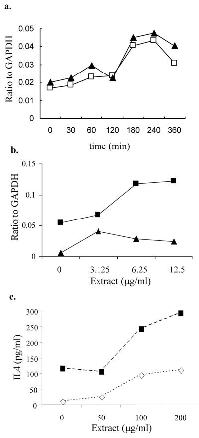
Figure 6.
Cytokine expression by mast cells exposed to cedar pollen extract.
a. RBL-2H3 cells were stimulated with 100 μg/ml cedar pollen extracts for 4 hours and IL-4 (open boxes; □) and IL-6 (filled triangles; ▲) mRNA relative to GAPDH was measured by RPA.
b. RBL-2H3 cells sensitized with anti-DNP IgE (500 ng/ml) were incubated for 4 hours with cedar extract alone (filled triangles; ▲) or with suboptimal concentrations of DNP-BSA (1.0 ng/ml) (filled boxes; □). IL-4 mRNA relative to GAPDH was measured by RPA. For reference, 50 ng/ml DNP-BSA stimulated an IL-4/GAPDH ratio of 0.1.
c. RBL-2H3 sensitized with anti-DNP-IgE were stimulated with pollen extract plus DNP-BSA at 1.0 ng/ml (◇; open diamonds) or 50 ng/ml (■; filled boxes). Culture supernatants were collected at 24 hours and analyzed for IL-4 by Bioplex. Culture supernatants from cells stimulate with pollen extract alone had undetectable levels of IL-4.

