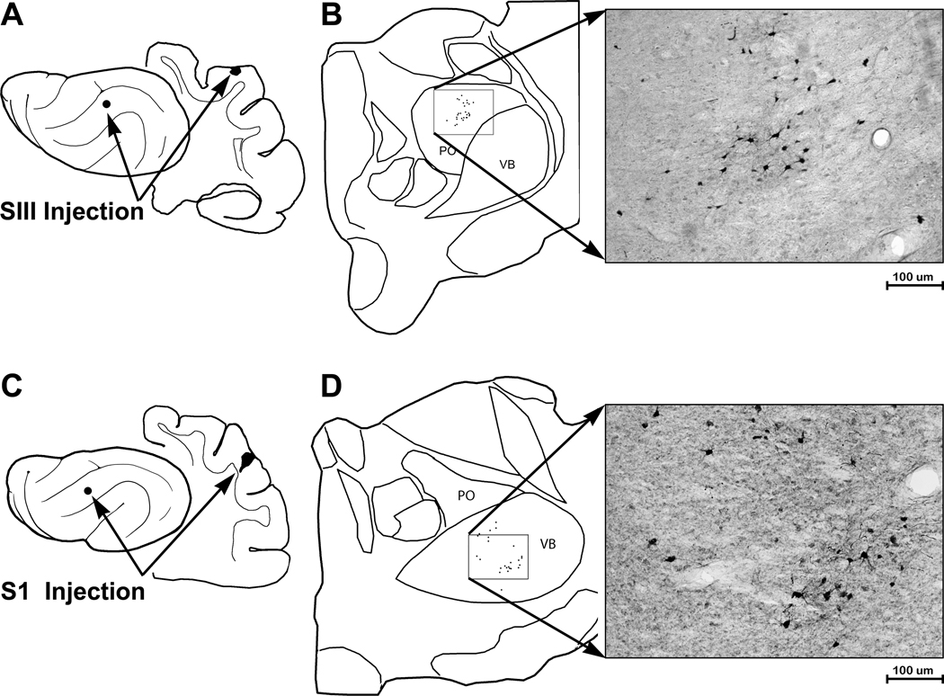
Figure 8.
Thalamic connections with SIII are different from those with S1. (A) The lateral view of the ferret cortex, and the coronal section, indicates the location of tracer injection into SIII. Part ‘B’ shows a tracing of thalamus with the subnuclei of posterior thalamic nucleus (PO) and ventrobasal nucleus (VB) outlined. For tracer injections into SIII, labeled neurons were identified and plotted in PO (boxed area), which is enlarged in the photomicrograph of labeled neurons in. Part ‘C’ illustrates the injection site in S1 on lateral and coronal views of the brain. Part ‘D’ shows a tracing of thalamus with the subnuclei of posterior thalamic nucleus (PO) and ventrobasal nucleus (VB) outlined. For tracer injections into SI, labeled neurons were identified in VB (boxed area), which is enlarged in the photomicrograph of labeled neurons.

