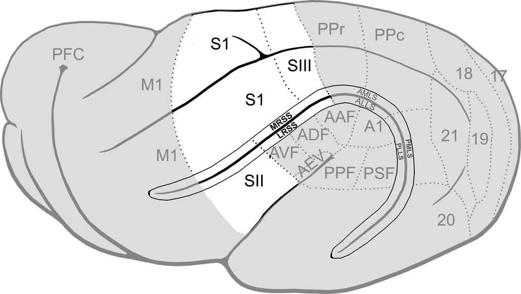
Figure 9. Summary of ferret cortical representations.
On this lateral view of the ferret cerebral cortex (left hemisphere; anterior=left), the known somatosensory regions are highlighted in white. The borders of functional subdivisions are represented by the dotted lines. Abbreviations: S1, somatosensory area 1; SII, somatosensory area 2; SIII, somatosensory area 3; MRSS, medial rostral suprasylvian sulcus; LRSS, lateral rostral suprasylvian sulcus.

