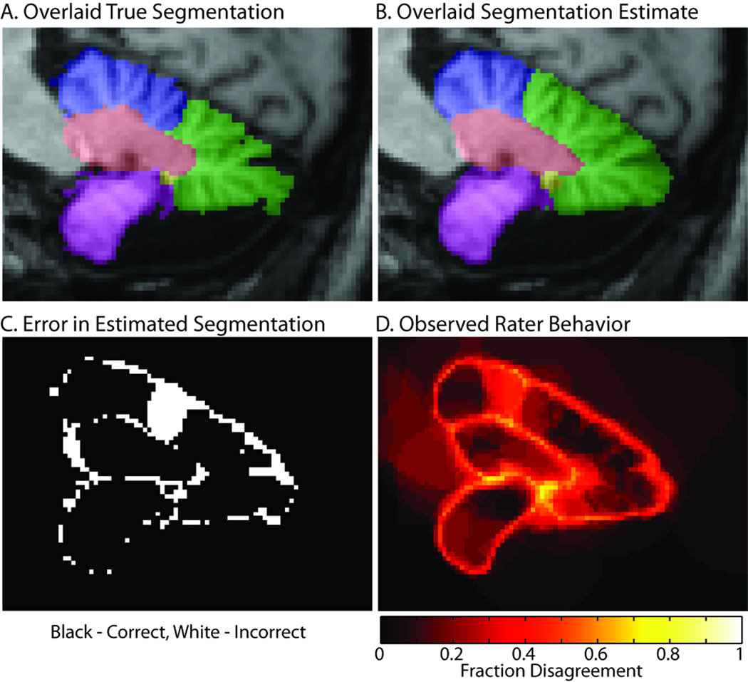
Figure 6.
Illustration of rater confusion. The expertly labeled truth dataset (A) has much greater detail in the cerebellar structures than is typical of a minimally trained rater, which tent to produce much smoother fused results (B). The errors (C) are largely concentrated around boundaries and result in smoothing and omission of fine division of minor sulci. However, there is a notable exception where the raters selected a different division between the superior (blue) and middle (green) lobules. Note that disagreements among raters (D) also occurred primarily along boundaries and in the region of error.

