Figure 1.

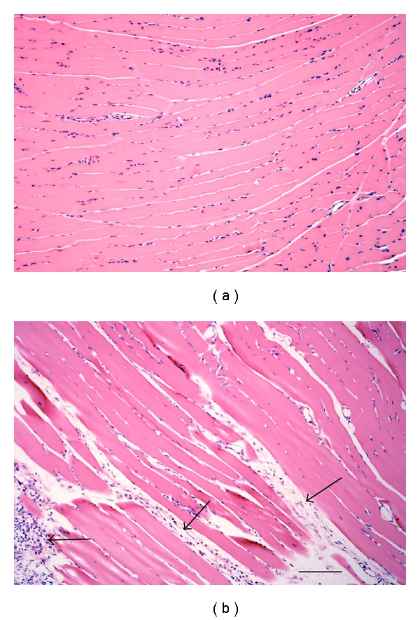
Photomicrograph of the masseter muscle stained with H&E. (a) Naive masseter muscle. (b) Masseter muscle 12 days after eccentric muscle contraction. Arrows indicate regions of fibrosis. Scale bar = 100 μm.

Photomicrograph of the masseter muscle stained with H&E. (a) Naive masseter muscle. (b) Masseter muscle 12 days after eccentric muscle contraction. Arrows indicate regions of fibrosis. Scale bar = 100 μm.