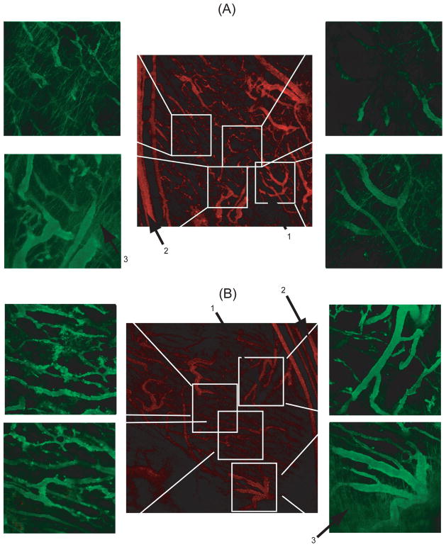
Figure 3.
Spatial distribution of (A) ELP-64/90 and (B) NGR-ELP-64/90 in FaDu tumor xenografts. ELP-64/90 did not show punctate fluorescence in the smaller central vessels and showed extravasation at the tumor periphery. NGR-ELP-64/90 showed punctate fluorescence in central vessels and extravasation at the tumor periphery. Arrow mark (1) smaller central vessels, (2) larger periphery vessels and (3) extravasation pattern.

