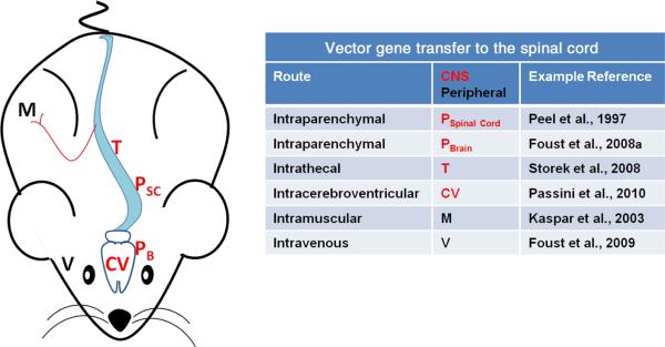
Fig. 2.
Routes of AAV vector administration for spinal cord transduction. CNS administrations target injections directly to CNS tissues or the cerebrospinal fluid (red). Intraparenchymal injections can be into spinal cord (PSC) tissue, or in brain tissues that have axonal projections into the spinal cord (PB). Injections can be into the intrathecal cavity (T) or the cerebral ventricles (CV), which can enhance vector spread in cerebrospinal fluid. Peripheral routes of administration lead to transduction of motor neurons: intramuscular (M) or intravenous (V) injections (black). Intravenous injections efficiently transduce neurons throughout the brain and spinal cord, although the approach is limited by having to use very young subjects. Modified vectors that can cross the mature blood–brain barrier (Gray et al., 2010) or the use of mannitol, which relaxes the blood–brain barrier (McCarty et al., 2009), could make this route more applicable in adults. Certainly some of these approaches transduce brain neurons in addition to spinal cord neurons (e.g. Foust et al., 2008a, 2009; Passini et al., 2010), although the figure highlights strategies to affect spinal cord diseases.

