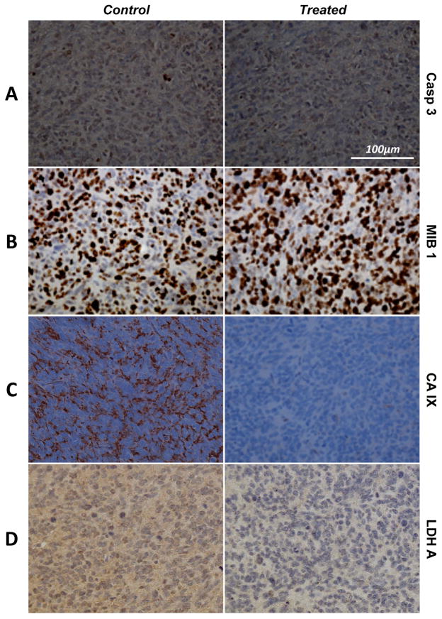
Figure 5.
(A) Casp3, (B) MIB-1, (C) CA-IX and (D) LDH-A IHC stains of orthotopic GBM tumors resected after 7 days of carrier (left, control) and Everolimus (right, treated) treatments. No differences are observed between the two samples for both Casp3 and MIB-1, suggesting identical levels of apoptosis and proliferation, respectively. However, a clear drop CA-IX and LDH-A can be seen in the treated tumor.

