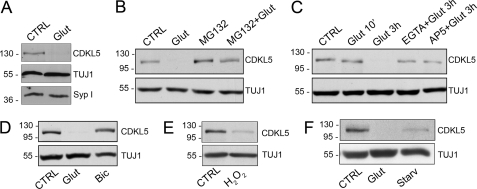
FIGURE 6.
A sustained extrasynaptic NMDA-R stimulation promotes CDKL5 degradation. A, Western blot analysis of CDKL5 and synaptophysin I (Syp I) expression levels in control neurons (CTRL) or after 3-h treatment with 10 μm glutamate (Glut). TUJ1 was used as loading control. B, immunoblot analysis of CDKL5 expression in unstimulated hippocampal neurons (CTRL) or in hippocampal neurons treated for 3 h with 10 μm glutamate (Glut), 50 μm MG132 (MG132), or both MG132 and glutamate (MG132 + Glut). β-Tubulin III (TUJ1) was used as internal standard. C, Western blot analysis of CDKL5 levels in hippocampal neurons treated for the indicated time with 10 μm glutamate alone or together with EGTA (2 mm) or AP5 (100 μm) compared with untreated neurons (CTRL). D, analysis of CDKL5 levels in untreated hippocampal neurons (CTRL) or treated for 3 h with either glutamate, 10 μm (Glut), or bicuculline, 40 μm (Bic). E, analysis of CDKL5 levels in neurons treated with H2O2 (50 μm) for 5 h (H2O2) compared with untreated cells (CTRL). F, analysis of CDKL5 levels in control neurons (CTRL) or in neurons treated with glutamate (10 μm) for 3 h (Glut) or starved for 24 h in neurobasal medium without B27 supplement (Starv).

