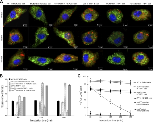
FIGURE 2.
Roles of the leptospiral InvA in invasion and survival in host cells. A, typical microphotographs of HEK293 and THP-1 cells co-incubated with the invA− mutant, invAcom revertant, and wild type of L. interrogans strain Lai for the indicated times. Blue: nuclei; green: cytoplasm; red: leptospires. B, fluorescence intensity reflects internalization levels of the invA− mutant, invAcom revertant, and wild type of L. interrogans strain Lai in infected HEK293 and THP-1 cells for the indicated times. Statistical data from experiments such as shown in A are shown. 100 cells were analyzed to quantify for each the values of fluorescence signal intensity. * and #: p < 0.05 versus both the wild type and invAcom revertant. C, colony-forming units of the intracellular leptospires recovered from THP-1 and HEK293 cells infected for the indicated times. * and #: p < 0.05 versus both the wild type and invAcom revertant.

