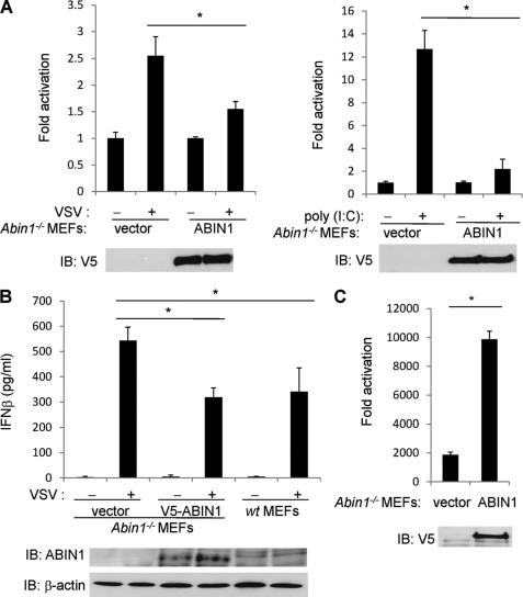
FIGURE 3.
Abin1−/− MEFs produce increased IFN-β in response to virus infection. A, Abin1−/− MEFs were transfected with IFN-β luciferase reporter (200 ng), pRL-tk (20 ng), V5-ABIN1 (1 μg) or empty vector (1 μg). Cells were then infected with VSV-ΔM (left) or transfected with poly(I:C) (12 μg) (right). IFN-β luciferase assays were performed 16 h later. B, Abin1−/− MEFs were transfected with either empty vector or V5-ABIN1 (0.3 μg of each). After 24 h, Abin1−/− and wild-type MEFs were infected with VSV-ΔM, and supernatants were used for an IFN-β ELISA 16 h later. The lysates were subjected to immunoblotting with anti-ABIN1 and anti-β-actin. C, Abin1−/− MEFs were transfected with pRL-tk (20 ng), V5-ABIN1 (1 μg), or empty vector (1 μg). Cells were then infected with VSV-luc, and IFN-β luciferase assays were performed 16 h later. *, p < 0.05. IB, immunoblotting. Error bars, S.D.

