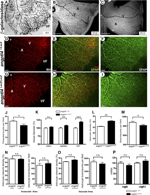
FIGURE 1.
Defective developmental angiogenesis in angptl4-deficient pups. A–C, LacZ staining shows expression of angptl4 in endothelial cells at P7 (A), P12 (B), and P17 (C) during developmental angiogenesis of the retina. Scale bar = 500 μm. D–F and G–I, IB4 (red) and GFAP (green) staining allow quantification of the vascular and astrocytic networks, respectively. A, artery; V, vein; VF, vascular front; o, optic nerve. Scale bar = 100 μm. Quantification of filopodia bursts/field of view (J); of artery, capillary and vein diameter (K); of vascular density (% of IB4 positive surface/total field area) (L); of vessel branch points/field of view (M); of astrocyte network density (left panel) and of astrocyte branch points/field of view (right panel) in the avascular (N) and vascular (O) area in angptl4+/LacZ and angptl4LacZ/LacZ P6 retinas (n = 8 per group). Shown is quantification by real-time quantitative PCR of vegfa and vegfr2 mRNAs (P) and in angptl4+/LacZ and angptl4LacZ/LacZ P7 retinas (n = 3 per group). GFAP, glial fibrillary acidic protein. ns, non significant.

