FIGURE 4.
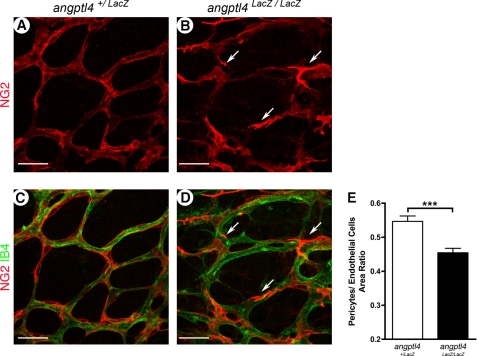
Defects in pericyte coverage in developmental angptl4LacZ/LacZretinas. A–E, delay of pericyte coverage in area 2 of P7 angptl4 LacZ/LacZ retinas. Representative images of P7 angptl4+/LacZ (A and C) and angptl4LacZ/LacZ (B and D) retinas stained with IB4 (red) and NG2 (green) (A–D). The arrows point to abnormal pericytes (B and D) on angptl4 LacZ/LacZ capillaries. Scale bar = 25 μm. E, shown is the ratio of pericytes (NG2+ area) to endothelial cell surface (IB4+ vessels), n = 6 per group. p < 0.005.

