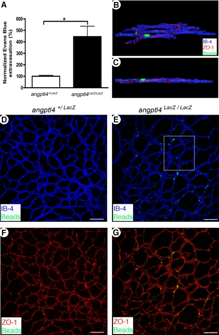
FIGURE 5.
Increased vessel permeability in angptl4-deficient pups. A, leakage of Evans Blue in P7 angptl4+/LacZ and angptl4LacZ/LacZ retinas. Values are expressed as % of the control (angptl4+/LacZ) value, (angptl4+/LacZ, n = 4; angptl4LacZ/LacZ, n = 5). B and G, confocal images of whole mount P7 angptl4+/LacZ and angptl4LacZ/LacZ retinas stained with IB4 (blue) and ZO-1 (red). Prior to sacrifice, fluorescent 100 nm microspheres were injected intravenously and flushed from circulation 15 min afterwards. B and C, deconvolution and three-dimensional reconstruction of the boxed region in E. Extravasated microspheres are trapped around tight junctions with a large part outside of the vessel (ZO-1, red). Field dimensions are 200 × 220 × 7 μm. E and G, extravasated microspheres (green) were only observed in angptl4LacZ/LacZ retinas. Scale bar = 100 μm. n = 6 per group. p < 0.05.

