FIGURE 4.
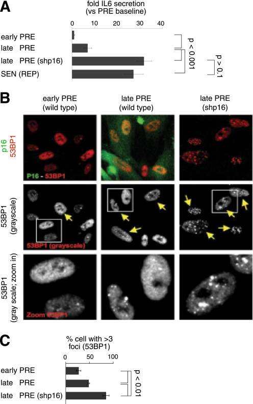
p16INK4a-depleted PRE cells develop early IL-6 secretion with 53BP1 foci formation. A, IL-6 secretion as a function of passage. CM from WI-38 cells infected with control (insertless) or shp16 lentivirus were analyzed for IL-6 by ELISA. We compared early (early PRE) and late (late PRE) passage cells with the same cells at complete replicative senescence (SEN(REP)). B, single cell analysis of p16INK4a and 53BP1 foci. p16INK4a and 53BP1 foci were detected by immunofluorescence. Upper panels show p16INK4a (green) and 53BP1 (red) co-staining. Middle and bottom panels show the grayscale for 53BP1 staining. The yellow arrows indicate cells with three or more 53BP1 foci. C, cell populations shown in B were analyzed for the percentage of nuclei positive for three or more 53BP1 foci. At least 200 nuclei were scored per condition.

