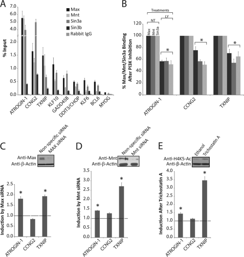
FIGURE 2.
Max, Mnt, and Sin3a bind to E-box sequences and repress transcription in proliferating cells. A, ChIPs were performed from proliferating T98G cells with anti-Max, anti-Mnt, anti-Sin3a, anti-Sin3b, and control rabbit IgG. Data represent the mean ± S.E. of 3 samples for Max, Mnt, and Sin3a, or 2 samples for Sin3b. Max, Mnt, and Sin3a binding at all E-box sites was significantly higher than the corresponding IgG controls (p < 0.05). B, ChIPs with Max, Mnt, and Sin3a antibodies from cells treated with either DMSO (NT) or LY294002 (LY) for 30 min. Data represent the percentage of binding after LY294002 compared with vehicle control. Data for Max are the mean ± S.E. of 2 and for Mnt and Sin3a the mean ± S.E. of 3 samples. Significant differences between control and LY294002-treated cells are designated (*, p < 0.05). C, actively proliferating T98G cells were transfected with Max siRNA. Data are presented as the fold-difference in expression between cells transfected with Max siRNA compared with nonspecific control siRNA and are the mean ± S.E. of 4 independent transfections. Knockdown of Max protein was 90–95% (see inset). Expression of ATROGIN-1 and TXNIP was significantly up-regulated after Max knockdown (*, p < 0.05). D, actively proliferating T98G cells were transfected with Mnt siRNA. Data are presented as the fold-difference in expression between cells transfected with Mnt siRNA compared with nonspecific control siRNA and are the mean ± S.E. of 4 independent transfections. Knockdown of Mnt protein was 60% (see inset). Expression of ATROGIN-1 and TXNIP was significantly up-regulated after Mnt knockdown (*, p < 0.05). E, proliferating T98G cells were treated for 6 h with trichostatin A to inhibit HDAC function. Expression of ATROGIN-1 and TXNIP was significantly up-regulated after trichostatin A treatment (*, p < 0.05).

