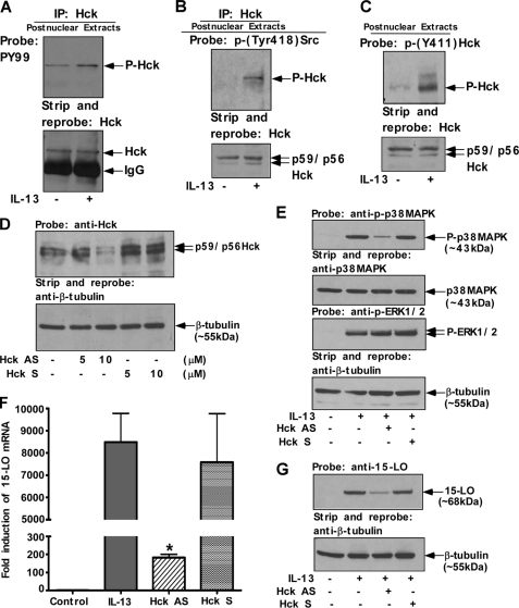
FIGURE 7.
Activation of Hck, the specific Src PTK isoform, regulates both p38 MAPK activation/phosphorylation and 15-LO gene expression in IL-13-stimulated monocytes. Monocytes (10 × 106/group) (A and B) and (5 × 10⁶/group) (C) were either incubated in medium alone or directly treated with IL-13 (2 nm) for 15 min. Postnuclear extracts were prepared and subjected to IP with Hck (goat polyclonal) antibody (A and B). The immunoprecipitates were resolved by SDS-PAGE for immunoblotting with the anti-phospho-tyrosine antibody, PY99 (mouse monoclonal) (A, upper panel). HRP-conjugated donkey anti-mouse, pre-absorbed secondary antibody from Affinity Bioreagents Inc. (Golden, CO), was used to develop the blot. In the upper panel of B, immunoprecipitates were resolved by 8% SDS-PAGE and immunoblotted with the rabbit polyclonal anti-phospho-Src (Tyr-418) antibody. HRP-conjugated donkey anti-rabbit, pre-absorbed antibody from Affinity Bioreagents Inc., was used as a secondary antibody. The blots were subsequently stripped and reprobed with Hck antibody to assess equal immunoprecipitation (lower panels of A and B). In the upper panel of C, postnuclear extracts (50 μg/lane) were resolved by 8% SDS-PAGE and immunoblotted with anti-phospho-Hck (Tyr-411) antibody (goat polyclonal). The blot was then stripped and reprobed with anti-Hck antibody as a loading control (lower panel). In D–G, monocytes (5 × 10⁶/group) were pre-treated with antisense (AS) or sense (S) ODNs to Hck either at indicated concentrations or at 10 μm for 48 h prior to the addition of IL-13 (1 nm) for either 15 min (E) or 24 h (F and G). D, cells were lysed and 50 μg of the postnuclear extracts (from each sample group) was separated by 8% SDS-PAGE and immunoblotted with anti-Hck antibody to examine the effect of antisense ODN on Hck expression (upper panel of D). The same blot was stripped and reprobed with an antibody against β-tubulin to assess equal loading (lower panel of D). In E, postnuclear extracts (50 μg/lane) were resolved by SDS-PAGE and immunoblotted with anti-phospho-p38 MAPK (Thr-180/Tyr-182) antibody. The blot was then stripped and reprobed with anti-p38 MAPK antibody. In another experiment, cells were lysed and 50 μg of the postnuclear extracts (from each sample group) was separated by 8% SDS-PAGE and immunoblotted with anti-phospho-ERK1/2 antibody. The same blot was stripped and reprobed with β-tubulin antibody to assess equal loading. The results are representative of three identical experiments performed. F, total cellular RNA extracts were prepared and subjected to real-time quantitative RT-PCR analysis. After normalization with GAPDH amplification, the -fold induction of 15-LO mRNA expression for different groups was plotted. Data are the means ± S.D. (n = 3). Significant differences were determined by comparing the antisense (AS) or sense (S) ODNs (to Hck)-treated groups to the IL-13-treated control (*, p < 0.004). In G, the cells were harvested and lysed. 50 μg of the postnuclear lysates was resolved by SDS-PAGE, and 15-LO protein expression was detected on Western blots with a 15-LO-specific antibody (upper panel of G). The 15-LO blot was stripped and reprobed with an antibody against β-tubulin (lower panel of G) to assess equal loading. The results are representative of three independent experiments.

