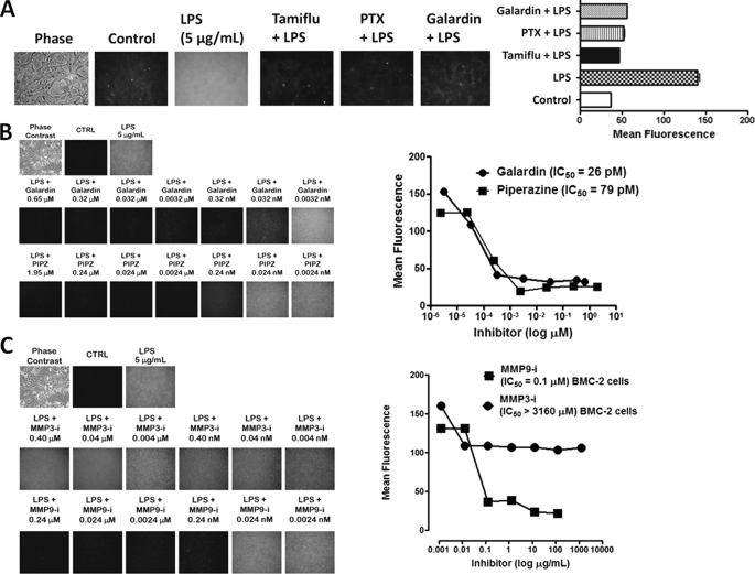
FIGURE 1.
A, LPS-induced sialidase activity is blocked by specific GPCR Gαi subunit and MMP inhibitors in live HEK-TLR4/MD2 cells. Cells were incubated on 12-mm circular glass slides in conditioned medium for 24 h at 37 °C in a humidified incubator. After removal of medium, 0.2 mm 4-MUNANA (4-MU) substrate in Tris-buffered saline, pH 7.4, was added with mounting medium to cells alone (Control), with 5 μg/ml LPS, or with LPS in combination with 250 μg/ml Tamiflu, 500 nm galardin, or 25 ng/ml PTX. Fluorescent images were taken at 1-min intervals using epifluorescent microscopy (×40 objective). The mean fluorescence surrounding the cells for each of the images was measured using ImageJ software. The data are a representation of one of six independent experiments showing similar results. Error bars, S.E. B, LPS-induced sialidase activity in live BMC-2 macrophages is inhibited by galardin and piperazine in a dose-dependent manner. After removing medium, 0.2 mm 4-MUNANA substrate in Tris-buffered saline, pH 7.4, was added to cells alone (CTRL), with 5 μg/ml LPS, or with LPS in combination with galardin (GM6001) or piperazine (PIPZ, MMP inhibitor II) at the indicated concentrations. Fluorescent images were taken at 1 min after adding substrate using epifluorescent microscopy (×40 objective). The IC50 of each compound was determined by plotting the decrease in sialidase activity against the log of the agent concentration. The data are a representation of one of three independent experiments showing similar results. C, MMP9i but not MMP3i blocks LPS-induced sialidase activity in BMC-2 macrophages. Images are as described in A. Data analyses are as described in B. The data are a representation of one of three independent experiments showing similar results.

