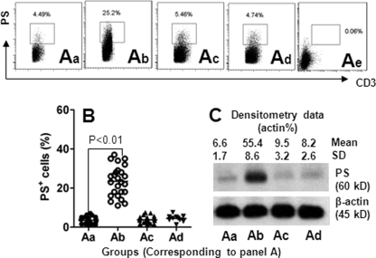
FIGURE 2.
Expression of PS in T cells is increased in glioma tissue. A, T cells (CD3+ cells) were isolated from marginal normal tissue (panel a; determined by pathologists), glioma tissue (panel b), and the peripheral blood of 25 patients with glioma (panel c), and the peripheral blood of 10 healthy volunteers (panel d). The cells were analyzed by flow cytometry and Western blotting. The dot plots in panels a–d show the frequency of PS+ CD3+ cells (gated cells). Panel e is an isotype control. B, scatter dot plots show individual data points of panels a–d in A. Each dot represents an individual data point. C, representative immunoblots (from 25 experiments) show IFNλ receptor protein levels from isolated T cells in panels a–d in A. Samples from patients were analyzed individually.

